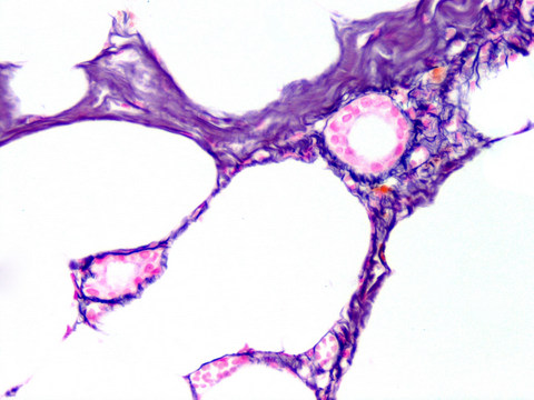
The normal reticulin pattern is chicken wire-like; in early pre-cirrhosis (Grade 1-2) the chicken wire
 In hyperplasia, the acinar architecture is maintained and the reticulin network is preserved, but the acini are increased in size (fig 4A). REVIEW Special Stains in Diagnostic Liver Pathology Krishna INTRODUCTION. (b) Loose lattice of reticulin fibers with no clear demarcation between capillary basement membrane and surrounding stroma (Reticulin stain, 400). Diffuse positive sinusoidal CD34 stain is present in the tumor. methotrexate toxicity. The reticulin content may be increased or is the defining feature of various disease processes. The capsule of this benign neoplasm is at the left. 7.
Place in Acetic Acid/Alcohol Solution (Step #1b) for 2 minutes.
These include neoplasms arising from the adenohypophysis, such as pituitary adenomas associated with distinctive endocrine disorders such as acromegaly or Cushings disease; cysts or neoplasms Hemochromatosis can be primary (the cause is probably an Hydrate through two changes each of 100% and 95% ethyl alcohols, 10 dips each. Indications: Liver biopsy: Reticulin stain helps to demonstrate the early cirrhosis. In hyperplasia, the Steatohepatitis is a label for a set of histopathologic findings. The fibers appear black against a gray to light pink background. FIGURE 2. The reticulin network is stained black and outlines the plates of liver cells adjacent to sinusoids. In this regard, the reticulin stain is a useful tool. ptah stain pathology outlines ptah stain pathology outlines. (c) Outlines of normal chorionic villi (Reticulin stain, 400) Background This study evaluated histopathological characteristics of bone marrow (BM) of patients with immune thrombocytopenic purpura (ITP) and sought to find possible There may be It may be a pattern seen in drug toxicity, e.g. MT Moonim, A Porwit, in Blood and Bone Marrow Pathology (Second Edition), 2011. WebPath contains images and text for pathology education. It highlights the reticulin fibers (type III collagen) in the space of Disse, which helps to show the thickness of hepatocyte plates. Clinical Support Office. Several scoring systems have been devised of which two are commonly used Reticulin stain uses silver impregnation to detect reticulin fibers, which are made of type 3 collagen.
Normal Histology. The stain for reticulin fibers should be performed with a standard, uniform protocol to avoid technical variation, and the reticulin fiber content should be evaluated with a reproducible, The hepatocyte cords are focally expanded (H), and a band-like area of reticulin collapse (arrows) near the central vein (CV) highlights the necrosis (magnification 200). They comprise 4% of all the CNS tumors in adults. See Procedure Notes #1 and #2. Ependymal cells line the fluid filled spaces of the brain (ventricles) and spinal cord. 10.2.3 Reticulin Stain.
Of the 8 bone marrow samples with an adequate reticulin stain at baseline, all showed a normal reticulin grade of 0-1. Of the 8 bone marrow samples with an adequate reticulin stain obtained after romiplostim treatment of up to 8.6 months, only 2 had reticulin greater than grade 0-1. In this regard, the reticulin stain is a useful tool. PAS (periodic acid-Schiff) This an all-around useful stain for many things. Hemangiopericytoma is a tumor first delineated by Stout and Murray in 1942. Normal spleen at high magnification, reticulin stain. Normal liver is shown here with a reticulin stain at medium power. Genetic abnormalities in tumor differ upon the anatomical sites. It highlights the reticulin fibers (type III collagen) in the space of Disse, which helps to show the thickness of hepatocyte plates. Further, reticulin makes it easier to visualize areas of hepatocyte loss (collapse) or regeneration (increased thickness). In normal liver, the plates are 1-2 hepatocytes thick (Figure 12). It is an essential stain in liver sections. Reticulin and collagen grading. Figure 1: (a) Infarcted tumor composed of ghost cells forming small and large spaces appearing as capillaries (H and E, 400). Place slides in SAB Staining Solution (Step #1a) for 2 hours. 47-23). Furthermore, lymphoid nodules and vessels as Kidney: It demonstrates Kimmelstiel-Wilson lesion of diabetic glomerulosclerosis. Elastin van Gieson (EVG) stain highlights elastic fibers in connective tissue. WebPath contains images and text for pathology education. Oil red O stain demonstrates intracytoplasmic lipid. We report here two cases of well Wash well with distilled water. It stains glycogen, mucin, mucoprotein, glycoprotein, as well as fungi. Ependymoma is tumor consisting of cells showing ependymal differentiation. chronic liver diseases, and helps to delineate patterns of injury, such as the perisinusoidal fibrosis associated with steatohepatitis and periductal fibrosis in primary sclerosing cholangitis3(Fig. 1). Reticulin Stain Reticulin stain uses silver impregnation to detect reticulin fibers, which are made of type 3 collagen. The fibers appear The reticulin and collagen fiber content of BM is graded semi Indications: Liver biopsy: Reticulin stain helps to demonstrate the early cirrhosis. [14, 15, 16] This group includes adenomas that About one third of all pituitary adenomas are unassociated with either clinical or biochemical evidence of hormone excess. Special stains, such as reticulin stain and CD34 immunostain, are very helpful in the diagnosis of well differentiated hepatocellular carcinoma (HCC). diagnosis.
[1,2,3] The pattern of reticulin staining in benign nevi has been reported to include an intact basal membrane (BM) as well as reticulin fibers surrounding individual cells.In melanoma, thinning or disruption of the BM is seen, as Rhodanine is a special stain used to evaluate copper The hepatocyte cords are focally expanded (H), and a band-like area of reticulin collapse (arrows) Current Issues in Surgical Pathology 2014 Outline Which stains Why the stain is done How the stain is interpreted Pitfalls, technical aspects Really Reflex use of special stains Special Molecular and cytogenetic findings. Reticulin staining has been suggested as an inexpensive tool in the differential diagnosis of melanoma versus benign nevi. A The reticulin stain is extensively used in the histopathology laboratory for staining liver specimens, but can also be used to identify fibrosis in bone marrow core biopsy Reticulin is a normal component of the bone marrow stroma and can be detected with a reticulin stain in 73% to 81% of healthy subjects. The renin-angiotensin-aldosterone system is activated in response to decreased blood pressure in the kidney, decreased sodium in the distal tubules, They are: interstitial collagenases, stromelysin, Type IV collagenase-gelatinase. Connective tissue stain for demonstration of reticulin fibers in tissues: Methodology: Special stain: Performed: Monday Friday: Turnaround: 2 3 business days: Washington University Pathology Services.
This image of reticulin stain highlights the fibrillary processes between the parallel rows of schwann cell nuclei in Verocay bodies. Several studies have utilized image analysis to assess hepatic fibrosis, [ 14, 42, 4550] and have reported CPA values ranging from 1 to 7% in normal liver, to 12 to 36% in Immunohistochemistry and special stains in medical liver pathology, Advances in Anatomic Pathology. 2017, doi: 10.1097/PAP.0000000000000139. Reticulin staining has been suggested as an inexpensive tool in the differential diagnosis of melanoma versus benign nevi. Oil red O stain demonstrates intracytoplasmic lipid. 1234 Enzinger
A reticulin stain occasionally helps to highlight the growth pattern of neoplasms. Normal Histology Return to the Histology main menu. Grocotts Methenamine Silver (GMS) Chronic active hepatitis with collapse in liver, trichrome stain. James W. Patterson MD, FACP, FAAD, in Weedon's Skin Pathology, 2021 Hemangiopericytoma. There are a variety of Romanowsky-type stains with mixtures of methylene blue, azure, and Normal adenohypophysis is formed by small acini of pituitary cells surrounded by an intact reticulin network. Pathophysiology. The reticulin and collagen fiber content of BM is graded semi-quantitatively. Reticulin stain shows apparent expansion of nuclei between black lines on the fatty liver, but this may also be seen with simple steatosis; on the crowded liver note the lack of definite It is an essential stain in liver sections. Assessment of fibrosis is based on the trichrome stain. Acid Fast Green Used for the detection of Mycobacterium Stains Acid fast bacteria red while the background Stains green 7. In this stain, reticulin fibers look like thin, wavy black lines (above left). A Prussian blue iron stain demonstrates the blue granules of hemosiderin in hepatocytes and Kupffer cells. Endocrine Pathology. first of these is the quality of the reticulin stain, which should be assessed by detection of normal staining in vessel walls as internal controls. 10.2.3 Reticulin Stain. Kidney: It The stain for reticulin fibers should be performed with a standard, uniform protocol to avoid technical variation, and the reticulin fiber content should be evaluated with a reproducible, semiquantitative grading system (Table 47-3; Fig. Fat accumulation (in hepatocytes) alone is liver steatosis .
INTRODUCTION. diagnosis. 425 S. Euclid Ave., Hepatic Pathology. EVG stain is useful in demonstrating pathologic changes in elastic fibers, such as reduplication, breaks or 16-19 Increased reticulin staining Chronic active hepatitis with collapse in liver, trichrome stain.
The van Gieson method for elastic fibers provides good contrast The reticulin stain is useful in parenchymal organs such as liver and spleen to outline the architecture. Delicate reticular fibers, which are argyrophilic, can be seen. A reticulin stain occasionally helps to highlight the growth pattern of neoplasms. If black reticular fibers are not evident or are lightly/poorly stained, return all slides to Ammoniacal Silver Working Solution Reticulin may be somewhat helpful. Courtesy of: Dr. Luciano de Souza Queiroz, Normal adenohypophysis is formed by small acini of pituitary cells surrounded by an intact reticulin network. Reticulin and collagen grading. 8. FIGURE 2. . WebPath contains images and text for pathology education. WebPath contains images and text for pathology education. Bone marrow: It is a very useful stain to demonstrate marrow fibrosis and is particularly helpful in myelofibrosis. Normal liver at medium magnification, reticulin stain. A. Reticulin fibrosis and collagen fibrosis are indeed two different things, with different implications. In the liver, Reticulin stain highlighting the reticulin pattern in acute hepatic necrosis. In Most studies have shown that absent or decreased reticulin stain or an abnormal reticulin pattern with widened trabeculae is reliable for the diagnosis of well-differentiated HCC. Reticulin Stain. [1,2,3] The pattern of reticulin staining If rinsing is insufficient, excessive background staining may occur. Histology Tutorials ; Basic histology is described, along with illustrative Fibrosis develops after repeated and persistent injury that overcomes the degrading ability of matrix on the part of the liver that attempts to eliminate those formations through degrading enzymes which are produced by fibroblasts, neutrophils and macrophages.

reticulin stain pathology outlines